
Discolored skin patches are irregular areas where there are changes in skin color. They are a common problem with a wide array of potential causes.
Some of the more common causes for changes in skin color are illness, injury, and inflammatory problems.
Discolored skin patches also commonly develop in a certain part of the body due to a difference in melanin levels. Melanin is the substance that provides color to the skin and protects it from the sun. When there is an overproduction of melanin in a given area, it can result in skin discoloration there.
Table of Contents
Here is a list of 18 possible causes.
Radiation therapy
- Only occurs in people being treated with radiation
- Blistering, dryness, itching, and peeling of the skin
- Hair loss at the site of treatment
Sunburn
- Superficial burn on the outermost layer of skin
- Redness, pain, and swelling
- Dry, peeling skin
- More severe, blistering burns may occur after extended periods of sun exposure
Candida
- Usually occurs in skin folds (armpits, buttocks, under breasts, between fingers and toes)
- Begins with itching, stinging, and burning red rash with wet appearance and dry crusting at the edges
- Progresses to cracked and sore skin with blisters and pustules that may become infected with bacteria
Rosacea
- Chronic skin disease that goes through cycles of fading and relapse
- Relapses may be triggered by spicy foods, alcoholic beverages, sunlight, stress, and the intestinal bacteria Helicobacter pylori
- There are four subtypes of rosacea encompassing a wide variety of symptoms
- Common symptoms include facial flushing, raised, red bumps, facial redness, skin dryness, and skin sensitivity
Burns
This condition is considered a medical emergency. Urgent care may be required.
- Burn severity is classified by both depth and size
- First-degree burns: minor swelling and dry, red, tender skin that turns white when pressure is applied
- Second-degree burns: very painful, clear, weeping blisters and skin that appears red or has variable, patchy coloration
- Third-degree burns: white or dark brown/tan in color, with leathery appearance and low or no sensitivity to touch
Tinea versicolor
- Slow-growing white, tan, brown, pink, or red spots on the skin that may be lighter or darker than your normal skin color
- Dry, flaky, and mildly itchy skin
- Skin areas that don’t tan
- Spots may disappear in cold weather and reappear in the spring and summer
Contact dermatitis
- Appears hours to days after contact with an allergen
- Rash has visible borders and appears where your skin touched the irritating substance
- Skin is itchy, red, scaly, or raw
- Blisters that weep, ooze, or become crusty
Strawberry nevus
- Red or purplish raised mark commonly located on the face, scalp, back, or chest
- Appears at birth or in very young children
- Gradually gets smaller or disappears as the child ages
Eczema
- Yellow or white scaly patches that flake off
- Affected areas may be red, itchy, greasy, or oily
- Hair loss may occur in the area with the rash
Bleeding into the skin
This condition is considered a medical emergency. Urgent care may be required.
- Occurs when a blood vessel bursts or leaks under the skin
- Bleeding into the skin can appear as small dots, called petechiae, or in larger, flat patches, called purpura
- The most common cause of bleeding under the skin is injury, but it may also be caused by more serious illness
- Always see a doctor about bleeding into the skin that isn’t related to a known injury, or if bleeding is causing excessive swelling or pain
Vitiligo
- Loss of pigment in the skin due to autoimmune destruction of the cells that give skin its color
- Focal pattern: loss of skin color in only a few small areas that may merge together
- Segmental pattern: depigmentation on one side of the body
- Premature graying of scalp and/or facial hair
Stasis ulcer
- Symptom of advanced stasis dermatitis
- Develop in areas of the body that have poor blood flow, most commonly in the feet and lower legs
- Painful, irregularly shaped, shallow wounds with crusting and weeping
- Poor healing
Basal cell carcinoma
- Raised, firm, and pale areas that may resemble a scar
- Dome-like, pink or red, shiny, and pearly areas that may have a sunk-in center, like a crater
- Visible blood vessels on the growth
- Easy bleeding or oozing wound that doesn’t seem to heal, or heals and then reappears
Actinic keratosis
- Typically less than 2 cm, or about the size of a pencil eraser
- Thick, scaly, or crusty skin patch
- Appears on parts of the body that receive a lot of sun exposure (hands, arms, face, scalp, and neck)
- Usually pink in color but can have a brown, tan, or gray base
Squamous cell carcinoma
- Often occurs in areas exposed to UV radiation, such as the face, ears, and back of the hands
- Scaly, reddish patch of skin progresses to a raised bump that continues to grow
- Growth that bleeds easily and doesn’t heal, or heals and then reappears
Melanoma
- The most serious form of skin cancer, more common in fair-skinned people
- Mole anywhere on the body that has irregularly shaped edges, asymmetrical shape, and multiple colors
- Mole that has changed color or gotten bigger over time
- Usually larger than a pencil eraser
Melasma
- Common skin condition that causes dark patches to appear on the face and, rarely, the neck, chest, or arms
- More common in pregnant women (chloasma) and individuals with darker skin color and heavy sun exposure
- No other symptoms beyond skin discoloration
- May go away on its own within a year or may become permanent
Mongolian blue spots
- Harmless skin condition seen at birth (birthmark)
- Most common in Asian neonates
- Large, flat, gray or blue patches with irregular edges seen on the back and buttock
- Usually fade away by adolescence

What causes discolored skin patches?
There are many potential causes of discolored skin patches, ranging from minor problems to more serious medical conditions.
Burns
Sunburns and other types of burns can damage your skin, and when these burns heal, there may be scar tissue that isn’t skin-colored. Discolored skin patches can also develop when you don’t apply sunscreen in a thorough manner, leading to a patchy tan. Certain medications can also make your skin more sensitive to the sun so that it’s more likely to turn red.
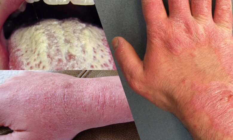
Infections
Various infections can cause localized changes in skin color. Cuts and scrapes may become infected when bacteria enter the wound, resulting in a skin infection. This leads to changes in the texture of the skin and turns the surrounding skin red or white. Fungal infections, such as ringworm, tinea versicolor, and candida can also trigger discolored skin patches on various parts of the body.
Autoimmune diseases and allergies
The immune system normally works to keep the body healthy by fighting off harmful invaders that cause infections and disease.
In people with autoimmune diseases and allergies, however, the immune system confuses healthy cells for something foreign and attacks them by mistake. This triggers inflammation throughout the body, resulting in various symptoms, including swelling and redness.
Some autoimmune diseases, such as lupus erythematosus and Graves’ disease, may attack the skin and cause changes in skin color. These reactions can range from red rashes and blisters to skin lightening or darkening.
Allergic reactions to foods, plants, or irritants can also result in discolored skin patches in various areas of the body. These changes may appear as rashes or raised bumps that itch or burn.
One common allergy that can cause skin discoloration is eczema. Like certain autoimmune diseases, eczema triggers an immune reaction that attacks the skin. The condition can cause scaly patches and red bumps that ooze or crust over.
Hormonal changes
Hormonal changes, especially during pregnancy, can trigger changes in skin color. These changes often occur due to increased levels of the female hormones estrogen and progesterone. Melasma, also known as the “mask of pregnancy,” is one skin condition that can develop due to these hormonal changes. It can cause dark patches to form on both sides of the face.
Birthmarks
Birthmarks are discolored skin spots that can develop at birth or after birth. Some common types of birthmarks include:
- Moles, which are brown or black spots that can appear on the skin at birth. Most moles aren’t cause for concern. However, changes in the size or shape of these spots can signal trouble and should be checked by your healthcare provider.
- Mongolian blue spots, which are bluish patches that can appear on the backs of babies and young children, usually those of Asian descent. They are harmless and often fade over time.
- Port-wine stains, which are flat patches that appear pink or red. They are caused by swollen blood vessels under the skin.
- Strawberry nevus, which is a red birthmark common in young children and infants. This birthmark usually goes away by age 10.
Skin cancer
Cancer can change the skin’s color or texture. Skin cancer may occur when the genetic material in skin cells becomes damaged, often from long-term sun damage or exposure to chemicals. The damage may cause the cells to grow out of control and form a mass of cancer cells.
There are several types of skin cancer, all requiring treatment:
- Actinic keratosis is a precancerous skin condition characterized by scaly, crusty spots on the hands, arms, or face. These spots are typically brown, gray, or pink. The affected area may itch or burn.
- Basal cell carcinoma is a form of cancer that affects the top layer of skin. It produces painful bumps that bleed in the early stages. The associated bumps may be discolored, shiny, or scar-like.
- Squamous cell carcinoma is a type of skin cancer that begins in the squamous cells. These cells make up the outermost layer of skin. The condition causes scaly, red patches and raised sores.
- Melanoma is the least common but most serious form of skin cancer. It begins as an atypical mole. Cancerous moles are often unsymmetrical, multicolored, and large. They usually first appear on the chest or back in men, and on the legs in women.
Most discolored skin patches aren’t caused by skin cancer. However, you should ask your healthcare provider to examine any misshapen moles or other rapidly changing skin lesions.
How are discolored skin patches evaluated?
You should schedule an appointment with your healthcare provider if:
- you have any lasting changes in your skin color
- you notice a new mole or growth on your skin
- an existing mole or growth has changed in size or appearance
Your healthcare provider will perform a physical examination and inspect your discolored skin patches. They will also ask you a series of questions about your skin changes. Be prepared to discuss:
- when you first noticed the change in skin color
- whether the discoloration happened slowly or quickly
- whether the discoloration is changing or getting worse
- any other symptoms you may be experiencing along with discolored skin
Make sure to notify your healthcare provider about any sunburns and other skin injuries. You should also tell your healthcare provider if you’re pregnant or taking any hormone treatments. These factors may play a role in your skin changes.
If your healthcare provider suspects that an underlying condition is causing your discolored skin patches, they will order certain diagnostic tests to pinpoint the cause. These tests may include:
- blood tests to check for conditions that may cause changes in skin color
- Wood’s lamp examination to identify possible fungal or bacterial infections
- skin biopsy to examine a small sample of the affected skin under a microscope for the presence of abnormal cells
How are discolored skin patches treated?
Treatment for discolored skin patches depends on the underlying cause. If your healthcare provider finds an underlying health condition, they will attempt to treat that particular condition first. The skin discoloration may be resolved with medical treatments or home remedies, or a combination of treatments.
Medical treatments
- Laser therapy: Intense pulsed light devices and Q-switched lasers are commonly used to help lighten skin areas that have darkened.
- Topical creams: Topical hydroquinone or prescription retinol (vitamin A) cream may help decrease the appearance of dark skin patches.
- Chemical peels: Chemical peels containing salicylic acid and glycolic acid can be used to remove the outer, discolored layer of skin.
Talk to your healthcare provider about your options so you can determine which treatment is best for you. Make sure to discuss the side effects, cost, and effectiveness of each treatment.
Home treatments
- Over-the-counter creams: Vitamin A cream or vitamin E cream can help reduce the appearance of skin discoloration and boost overall skin health.
- Lemon juice: Apply lemon juice twice per day to lighten skin areas that have darkened. This may reduce the appearance of discolored skin patches in six to eight weeks.
- Castor oil: Apply castor oil to discolored areas twice per day, or wear a bandage soaked in castor oil overnight. This can help smooth the skin and break down excess melanin.
- Vitamin C: Eat foods rich in vitamin C, an essential nutrient for skin health. Fruits high in vitamin C include cantaloupe, oranges, and pineapple.
- Drink tea: Drinking tea made from burdock, red clover, or milk thistle might reduce skin discoloration.
For any important information please contact us Email GadgetsNg info@gadgetsng.com
[Button id="1"]


Let’s spread the love! Tag a friend who would appreciate this post as much as you did.
I appreciate your creativity and the effort you put into every post. Keep up the great work!
I don’t think the title of your article matches the content lol. Just kidding, mainly because I had some doubts after reading the article. https://accounts.binance.com/bn/register-person?ref=UM6SMJM3